
By David Hudnall, DMD
Although most methodologies involving digital dental technology are roughly less than 25 years old, the beginning of the digital dentistry revolution seems like it was light years ago. That is because digitalization was a culmination of many independent factors that coalesced and allowed technology to develop by leaps and bounds within a few short years.
While we dental professionals may like to believe that digital dental technology has reached its pinnacle, the truth is there are more changes and innovations on the way.
A Pre-Digitization World
Prior to the development of digital dental technology, visualization and 2D X-rays were used to diagnose and plan treatment cases. Fabrication of both fixed and removable restorations was accomplished using the error-prone analog lost wax technique. The field of orthodontics was limited to traditional bands, brackets, and wires. Implant dentistry was practiced by eye-balling the best location for an implant without ever being completely certain what structures may be within the path of insertion.
While each of these methods clearly had merit and offered a history of proven successes, accuracy, and precision were often missing from the oral health equation. As dental digitalization began to provide clinicians with more information, it became possible to do more things that truly help our patients.
Advanced Digital Dental Technology
Most innovation in digital dentistry is currently focused on improving digital dental technology that provides enhanced diagnostics and treatment planning capabilities or automation that enhances dental outcomes and patient experiences.
Cone-Beam Computed Tomography (CBCT)
Similar to traditional planar radiography, 3D imaging has undergone a tremendous evolution since its inception for dental use in 1997. CBCT uses a cone- or pyramid-shaped x-ray beam in conjunction with a 2D detector array to obtain a large series of projections from the scanned objects that are reconstructed into a stack of 3D images.
Advancements in hardware technology and improved software capabilities have resulted in a range of current and future opportunities for optimizing CBCT imaging in digital dental technology. Let’s discuss a few of the technologies and projects currently in the research and development phase.
Longevity of the X-Ray Tube
The focal spot is one of the key elements that determines the resolution of an X-ray image. Currently, the typical nominal focal spot size in dental CBCT is 0.5 mm. This small focal spot enables high-resolution imaging but can result in overheating and damage to the tube anode.
While overheating is currently minimized by using relatively low exposure times, further refinements to X-ray tube technology will result in even smaller focal spots with less potential for damage to the equipment.
Beam and Rotation Geometry
A special technique that is starting to be introduced to the market is the use of a Reuleaux triangular-shaped field of view (FOV), which corresponds more closely to the shape of the teeth and dental arches compared to the cylindrical or spherical FOV options in current use. This change would lead to an even further reduction in radiation dose for scans of the dental region.
Reconstruction
Other potential improvements to CBCT that are gradually being introduced into practice are the use of metal artifact reduction algorithms and methods to correct patient movement. Both allow for improved image sharpness and images of greater diagnostic quality.
Optical Imaging
Finally, the ability to use open-source software to combine intraoral scanning data with CBCT images will allow complementary information from both types of scans to be utilized together for improved patient treatment planning and optimal patient comfort in prosthetic and implant cases.
Computer-Aided Designing (CAD)
While the history of CAD closely parallels the history of computers in general, CAD was developed decades ahead of portable computers that would eventually make the software available to anyone. CAD software evolved over the years, with the manufacturing sector making use of its design capabilities to create new products with precise measurements and tolerances.
This technology was borrowed and further adapted for digital dental technologies in use. Current CAD systems used in digital dental technology have transformed prosthetic restorations from an art form to a science, allowing dental technicians to conceive and produce restorations of ideal thickness and dimensions for improved structural integrity, stability, and longevity.
The future of CAD technology is continuously evolving, and several trends are shaping its trajectory. Some of the innovations on the horizon include cloud-based CAD systems that allow sharing of designs with multiple users across multiple devices and locations and designs that take advantage of algorithms and artificial intelligence (AI) to explore numerous design possibilities to determine the best solution based on specified parameters.
AI and machine learning will further automate digital technologies, providing design recommendations based on historical data and user preferences. Finally, CAD will focus on user-friendliness and intuitive design for enhanced productivity and a more enjoyable user experience.
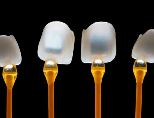
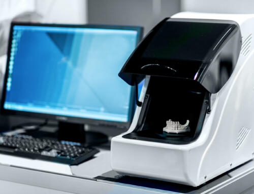
Intraoral Scanner
The intraoral scanner (IOS) is a device used for capturing impression data employed in the fabrication of a whole series of prosthetic restorations, orthodontic appliances, guided surgical templates, custom functional appliances, and more. The availability of chairside intraoral scanners equipment offers numerous advantages over traditional impression methods including speed, convenience, digital data capture with seamless integration into the digital dental laboratory workflow, and patient acceptance.
Still, researchers are focused on limitations and developing methods to improve digital impressions technology by incorporating artificial intelligence (AI) and cutting-edge software to make the devices more user-friendly and easier to integrate with other hardware.
Currently, the most common universal file format for 3D printing is STL. It was released in 1987, and it is very basic. An STL file describes the raw, unstructured, triangulated surface of the scanned object. It contains no scale, color, or material information. It contains no validation mechanism, so it doesn’t prevent operator error. While it’s good enough in most cases, it will be replaced shortly by 3D manufacturing format (3MF), a free, open-source code being developed by a consortium from a variety of different industries specifically designed for additive manufacturing, like 3D printing.
3MF files contain the actual 3D model and associated data that eliminate ambiguity associated with STL files while allowing your models to be saved and shared without machine-dependent software manipulation that consumes time.
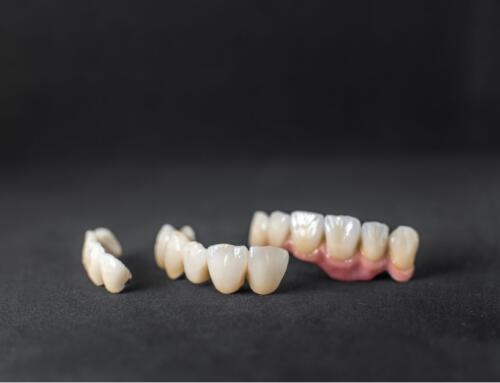
Computer-Aided Manufacturing (CAM)
Prior to CAM, manufacturing was a manual process that relied on the skill of artisans for production. It was time-consuming, labor-intensive, and error-prone. The development of computer-aided design (CAD) ushered in the possibility of creating detailed, precise digital models that generate instructions for additive or subtractive automated manufacturing.
The CAM software begins with the digital model from the design software and creates a series of instructions that allow a useful physical object to be produced. In the case of dentistry, this is usually some type of restoration or dental appliance.
- Improvements to CAM often revolve around finding solutions in technology that currently place limitations on product creation. In dentistry, that may be the raw materials themselves, the 3D printer, or the milling machine. Innovations currently under investigation include:
- Methods to decrease manufacturing time while maintaining the quality of the completed dental device.
- Addressing cross-contamination.
- Prevention of residual chemicals from being transferred to the patient.
- Determining methods to reliably 3D print permanent dental restorations from materials other than resins.
Digital Dental Technology with Stomadent Dental Lab
While it is certainly possible to own in-office 3D printers and milling machines, unless you utilize them every day, they are nothing more than expensive gadgets collecting dust. Why not pay for only the services that you actually use by taking advantage of the CAD/CAM digital workflows and expertise offered by Stomadent Dental Lab?
From single crowns to implant-supported full mouth restorations, we have you covered. Let Stomadent be a remote extension of your dental team with our innovative digital dentistry technologies!
Related Posts
![Hybrid Denture with Titanium Bar [Best Methods + Advantages]](https://stomadentlab.com/wp-content/uploads/2024/01/dental-prosthesis-on-dark-background-2023-11-27-05-06-28-utc-scaled-500x383.jpg)
![The Lucitone Denture Advantage [Best Practices + Advice]](https://stomadentlab.com/wp-content/uploads/2022/08/lucitone-promo-1-500x383.jpg)
![How to Remove Snap On Dentures [Expert Guidance]](https://stomadentlab.com/wp-content/uploads/2023/12/a-denture-in-a-glass-of-water-dental-prosthesis-c-2023-11-27-04-50-54-utc-scaled-500x383.jpg)


Request a FREE Dental Lab Kit