
By David Hudnall, DMD
The concept of minimally invasive dentistry is not new. Preserving as much good tooth structure is always preferable to replacing natural enamel and dentin with artificial substitutes that have a limited lifespan. Until recently, methodology did not exist to allow clinicians to accurately locate and diagnose caries in its infancy.
Tooth decay is almost always more advanced than a standard periapical or bitewing radiograph would indicate. Technological advances have allowed dentists to detect dental decay earlier and pinpoint its exact location much more precisely, reducing guesswork and allowing for more conservative treatment.
What Is Minimally Invasive Dentistry?
Minimally invasive dentistry emphasizes the preservation aspect of maintaining healthy tooth tissue during restorative treatment by using the least amount of dental intervention possible. It focuses on disease prevention, remineralization, and early detection of tooth decay. With minimally invasive dentistry, dentists use advanced techniques such as magnification, transillumination, imaging, and caries indicators to determine the location of caries and remove decay while preserving as much sound tooth structure as possible.
Minimally invasive dentistry is also sometimes referred to as micro-dentistry, which is designed around the concept that keeping natural dental tissue intact is better for the patient than replacing good tooth structure with a non-biological restorative substitute.
Benefits of Minimally Invasive Dentistry
Traditional techniques utilized in restorative dentistry often involve removing sound tooth structure in order to access the decay. This is particularly evident when interproximal decay is addressed by cutting through healthy tooth structure until the decay is located. Good tooth structure is further removed using the G. V. Black “extension for prevention” concept, which allows space for bulky matrix placement and a large quantity of restorative material.
Newer detection methods have advantages over radiographs that only allow the clinician to view the location of decay buccolingually. By having more information and a better understanding where the decay is located allows the dentist to make a more conservative cavity preparation, minimizing the need to guess or further explore in order to find the tooth decay.
Compared to past generations, a greater percentage of people aged 50 and over are able to maintain most of their natural teeth for their entire life. Therefore, it is critical to detect tooth decay early, before it has had the opportunity to advance and destroy vital tooth structure. When decay is caught early, less invasive treatment can be performed so that patients can maintain a greater quantity of natural tooth structure for the rest of their lives.
Minimally invasive dentistry is the antithesis of “extension for prevention”. Although large restorations have their place when decay is advanced, maintaining more healthy tooth structure translates into
- less occlusal loading and stress placed on restorations,
- longer lasting restorative materials,
- smaller preparations necessitating less time and artificial materials,
- stronger teeth,
- and reduced patient discomfort.
Techniques for Minimally Invasive Dentistry
The ultimate goal of minimally invasive dentistry is to extend the life of any restored teeth with as little intervention as possible. This means that we must use accurate diagnostic techniques, the best isolation and preparation techniques possible, and restorative materials that, with good oral hygiene, will last for many years.
Let’s discuss some of the important considerations when employing minimally invasive dentistry techniques to treat tooth decay.
Advanced Preparation Design
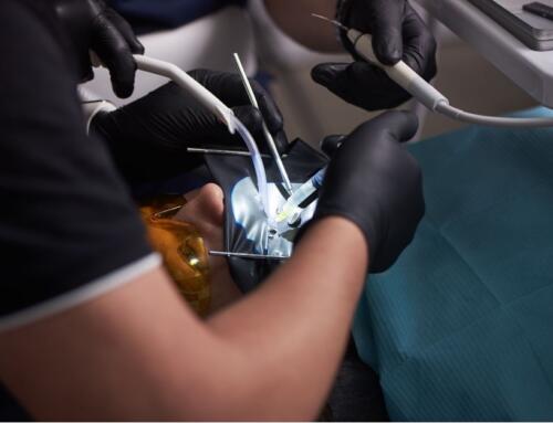
Because of being more certain of the location of caries in all directions, it is possible to make a smaller opening to access and remove decay located near fissures or near the interproximal contact without aggressive removal of excess tooth structure. Access to small lesions is often better facilitated through the use of a pediatric handpiece that has a smaller head design that improves visualization.
With the advent of injectable restorative materials, it is possible to reliably fill small cavity preparations that possess excellent margins and are free of voids, limiting exposure to harmful bacteria and risk.
The Canary System
Quantum Dental Technologies has developed a diagnostic device called the Canary System. According to the manufacturer, “The Canary System is a low-powered laser that facilitates early detection of dental caries and defects within the crystalline structure of the enamel before it is discernible on radiographs or visible clinically. It is able to detect decay within smooth enamel surfaces, on root surfaces, occlusal surfaces, interproximal regions, under dental sealants, and around existing amalgam or composite restorations.”
The system can detect caries as small as 50 microns up to 5 mm below the tooth surface. It is also useful in the detection and diagnosis of enamel cracks.
In addition to a digital photograph of the lesion with surrounding tooth structure displayed on a screen, a Canary exam features both audible and visual numeric measurements that help patients understand the rationale and benefits of early intervention via remineralization therapy or restorative treatment using minimally invasive dentistry. The Canary number is higher as the size of the lesion increases. Readings taken of the same location after remineralization or restorative treatment show a lower number when compared with the pre-treatment reading.
Modern Digital Radiograph Evaluation
When you think about it, modern digital radiology incorporates seamlessly with the concept of minimally invasive dentistry. After all, digital radiographs provide more information than traditional film with much less patient exposure to radiation. Digital radiography also allows images to be magnified and viewed by enhancing parameters, including
- contrast,
- colorizing,
- 3-D,
- sharpness,
- and zoom
To assist in the detection and interpretation of diagnosis. Digital images of problem areas are easily transferred and projected onto a computer screen in the operatory for patient viewing and education.
Metal Shim Devices
Ultra-thin metal shims, such as the Fender Mate system, are perfectly suited for addressing and restoring interproximal caries in the most conservative manner possible. After tooth preparation, the metal shim device with its preformed contact dimple is wedged between neighboring teeth and aids the clinician in rebuilding the contact point.
Because tooth preparation is minimal and isolated to the contact zone, the moldable metal shim serves as a matrix to support injectable composite resin during placement. Once the restorative material is cured and the shim is removed, the teeth will spring back into position via their respective periodontal ligaments, and the contact will become tight.
Interproximal Guard
When an interproximal tooth preparation is necessary, the worst thing you can do is to cause iatrogenic trauma or damage to an adjacent tooth or contact zone. You simply can’t put back the smooth surface of virgin enamel. Using an interproximal guard, such as Wedge Guard brand or Fender Wedge brand, can protect teeth against inadvertent bur strikes by putting an interproximal wall of metal between your bur and the adjacent tooth structure.
After preparation, the guard may be removed and replaced with a wedged metal shim device (discussed above) to ensure restoration placement is addressed properly. Interproximal guards may also be used when preparing teeth for indirect restorations.
Stomadent for Minimally Invasive Dentistry
There is no substitute for preserving solid, vital tooth structure. Stomadent Dental Lab understands that often the best dental treatment is no treatment at all. When treatment is indicated, conservative is better.
However, if more than 50% of the tooth is no longer sound, dental crowns are usually the restoration of choice. We offer digitally-designed and milled crowns from a variety of durable materials to address your patient’s every need. Explore the benefits of CAD/CAM-produced restorations to see all of the advantages that digital dentistry has to offer.
Related Posts
![Dental AI [The Latest Innovations in Clinical AI]](https://stomadentlab.com/wp-content/uploads/2024/04/patient-and-dentist-looking-at-results-in-a-screen-2023-11-27-05-23-38-utc-scaled-500x383.jpg)


Request a FREE Dental Lab Kit